Complete visual examinations
A complete visual eye examination, especially if performed periodically, is the best way to prevent at an early stage any visual defect or pathology that may compromise our vision. Apart from detecting the most common visual defects such as myopia, hyperopia, astigmatism and presbyopia among others, it may be the only way to diagnose in time really important diseases that can cause irreversible vision loss and, in more extreme cases, even blindness.
What does a complete visual examination consist of?
First, before proceeding with the tests, the optometrist will ask several questions about lifestyle, hobbies, possible diseases that may affect vision, ocular histology and family history of eye diseases. This information is of vital importance to know the special needs of each patient.
Use of the contact tonometer
Tonometry measures the pressure inside the eye. During the examination, the optometrist will use an instrument called a tonometer to measure eye pressure.
This test is complementary to detect glaucoma. Most cases of glaucoma are diagnosed with a pressure exceeding 21 mm Hg. However, some people may have glaucoma with pressures between 12 and 22 mm Hg and others may not have glaucoma with pressures above these measurements. Eye pressure is unique to each individual and further testing is always necessary to make a diagnosis.
The autorefractometer-keratometer.
The autorefractometer-keratometer electronically measures the possible refractive defects of the eyes and provides the optometrist with an approximate reading of the graduation, as well as the ocular parameters necessary for the adaptation of contact lenses, in this way the examination will be carried out with more accuracy and lightness.
The retinoscope
It is a high-precision instrument used to check the eyesight of very young children or people with communication difficulties who find it difficult to describe how they see. It replaces the autorefractometer in these cases.
In children, other tests such as steriopsis (binocular fusion test), the Ishihara test (to detect color blindness) and the eight gaze positions test (for the exploration of eye movements) are also added.
Subjective examination.
To refine the results, the optometrist will ask you to read the optotypes (letters, numbers or drawings in the case of the youngest children) and to answer questions about different tests with figures and symbols, first in monocular (with one eye) and then in binocular (with both eyes). Depending on the answers, the optometrist refines the prescription until the patient sees as clearly and comfortably as possible by adjusting the lenses in front of the eyes with the frontophocometer or trial spectacle.
Next, the necessary tests are performed to ensure good binocular vision and, only if necessary, near addition is measured.
Cover Test and Fixation Disparity.
If any binocular anomaly has been detected previously in the visual examination, these tests are used to determine the value of the angle of deviation of the visual axis at distance and near and to correct it, if possible, with prisms and/or vision therapy.
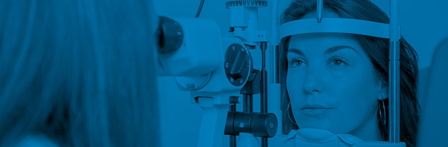
The slit lamp
Slit lamp examination is a magnified analysis of the eye from front to back. The head rests on equipment called a slit lamp and the eye care professional combines a bright light with different magnifying lenses to view the structure of the eye from the eyelids to the retina (the latter with the aid of a VOLK +90D lens).
A slit lamp is one of the most common procedures in a comprehensive eye exam because it provides a very complete report on eye health and uncovers indicators of many diseases and conditions, including dry eye, various complications from contact lens misuse, infections and alterations in the anterior segment of the eye, cataracts, glaucoma, diabetic eye, macular degeneration, and even blood disorders or cancers.
It is also a fundamental test for contact lens fittings.
Use of Retinoscopy
The retinoscope, like the VOLK +90D lens in the slit lamp and the ophthalmoscope, is used to examine the back of the eyeball with a digital photograph showing the entire retina, blood vessels, macula and the entrance of the optic nerve. This important test detects changes that indicate the presence of diseases such as glaucoma, diabetic eye, macular degeneration and even blood disorders and cancers.
This test is performed in a darkened room.
Final diagnosis.
At Interoptics we take it very seriously to convey and explain the different needs of each patient while attending to all their doubts and questions. The optometrist will take the time to explain the results of the tests and will advise you at all times with only the best interests of your eye health in mind.
Final advice.
For us, the final assessment is just as important as the complete visual examination. If necessary, a professional will guide you from choosing the eye care products that best suit your needs, to choosing a frame suitable for your personal comfort and convenience, to the lenses that are right for you. We want your visit to Interoptics to always be a good experience.


Free eye exam:
Conditions:
We perform one free check-up per person per year, if the patient needs to bring a report with his graduation but does not need to bring a report with his graduation.
If you do not wish to purchase any product, a charge of 20 euros will be applied. If you need a detailed report with the addition of retinography and
PIO, a charge of 30 euros will be applied.